|
||
|
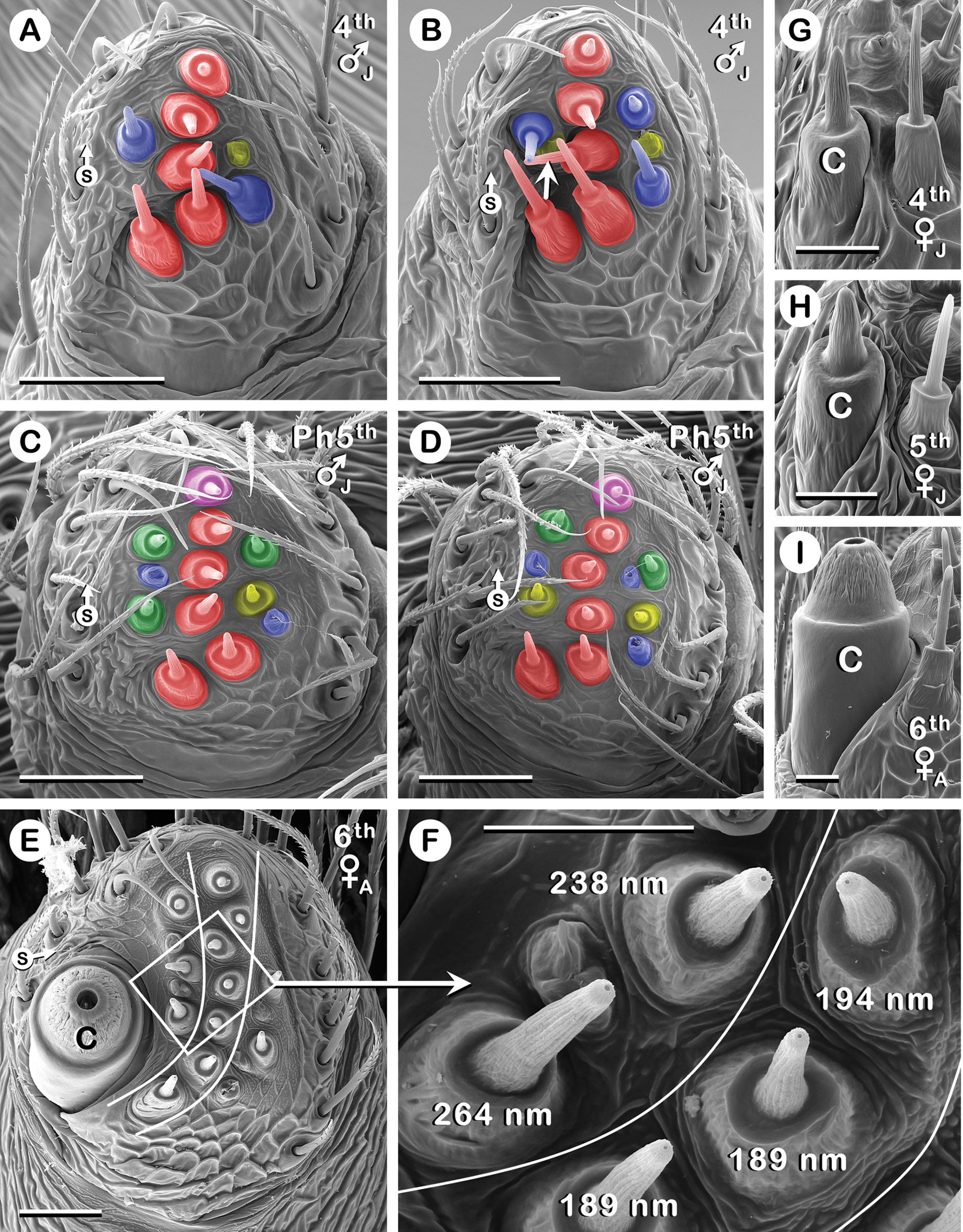
PLS of 4th to 6th instars of Australomimetus spinosus. A–D.PLS from one juvenile specimen, well into proecdysis. Color coding explained in Results (Convention 5 in ‘Conventions applied…’). A, B. Old exoskeleton. C, D. New exoskeleton directly beneath (A, B), respectively. One of two AC tartipores (yellow) in (B) largely obscured (unlabeled arrow). E, F. Adult, AC spigots between two white curves are non-T-A; the rest, anterior and posterior, are T-A. AC spigots in boxed region of (E) magnified in (F), showing larger diameter openings in T-AAC spigots compared with neighboring non-T-AAC spigots [see Results (‘PLS’)]; measurements given in nm [see Materials and methods (‘SEM of spinnerets…’)]. G-I. Ontogenetic changes in CY spigot (C) morphology; medial, high tilt view. Note changes in width relative to AC spigot on right. (E, F, I) all from same PLS. B, D–F, I. Left PLS. A, C, G, H. Right PLS (image flipped). C, CY spigot; S, putative sensillum. Scale bars: A-E 20 µm; F-I 10 µm. See also Abbreviations and terminology (‘Spinning apparatus abbreviations’) and Results (Convention 3 in ‘Conventions applied…’). |