|
||
|
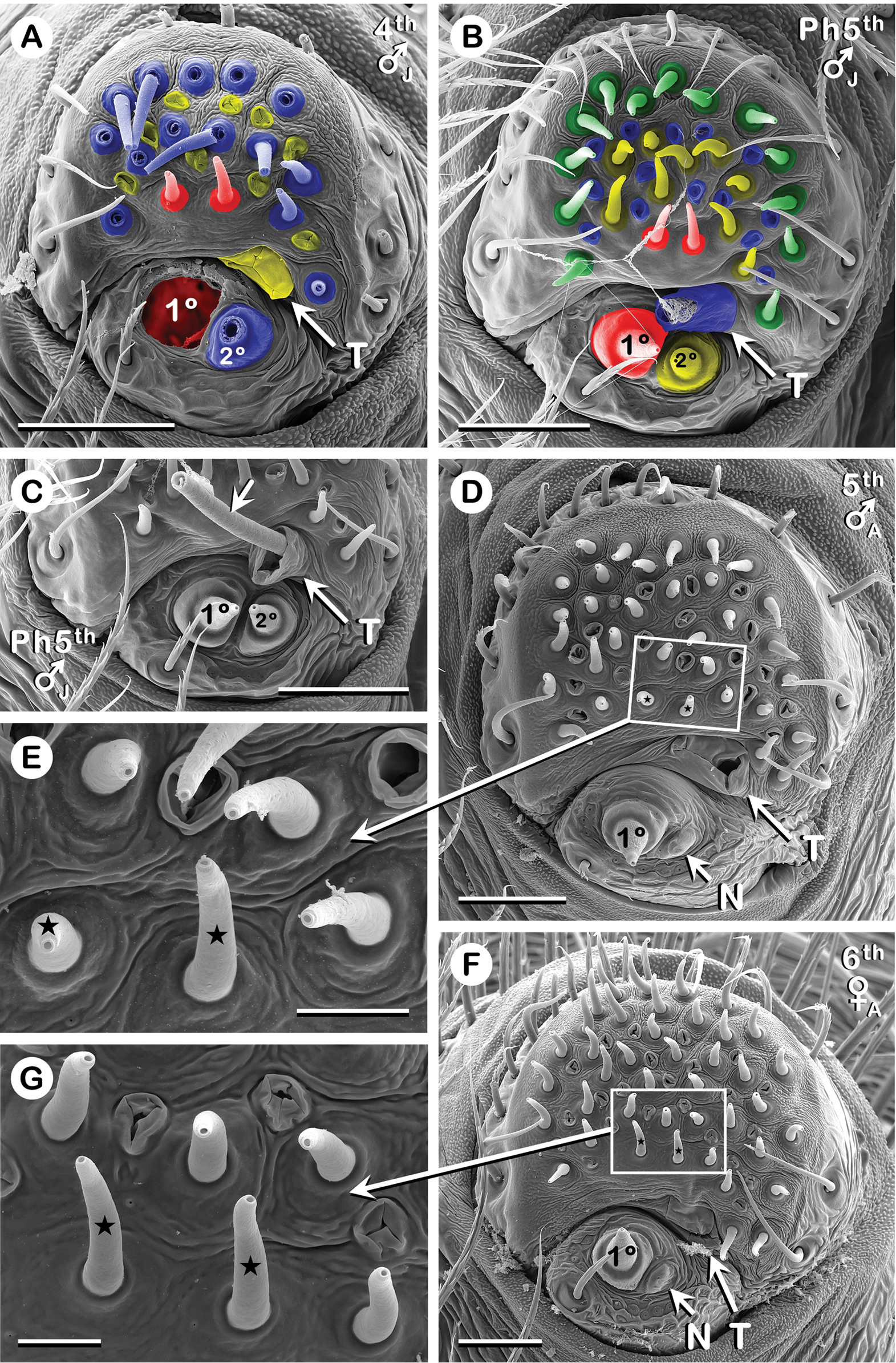
ALS of 4th to 6th instars of Australomimetus spinosus. A-C.ALS from one juvenile specimen, well into proecdysis. Color coding explained in Results (Convention 5 in ‘Conventions applied…’). A. Old exoskeleton (all spigots but one damaged; 1° MaA spigot torn out entirely, but its former location is colored red). B. New exoskeleton directly beneath (A). C. New exoskeleton, opposite ALS from (A, B), demonstrating utility of a tartipore. Unlabeled arrow to 2° MaA duct (severed at distal end) still accommodated by 2° MaA tartipore (T). Before old and new exoskeletons were separated, this duct emptied on a 2° MaA spigot on old exoskeleton (like that in (A) but on opposite ALS). D-G. Adults. Presumed pair of non-T-API spigots (stars) on each ALS in males (D, E) were morphologically similar to T-API spigots (unlabeled PI spigots) in both sexes and to non-T-API spigots (stars) in females (F, G): no MoPI spigots. C. Left ALS. A, B, D-G. Right ALS (image flipped). 1°, 1° MaA spigot; 2°, 2° MaA spigot; N, 2° MaA nubbin (ontogenetic); T, 2° MaA tartipore. Scale bars: A-D, F 20 µm; E, G 5 µm. See also Abbreviations and terminology (‘Spinning apparatus abbreviations’) and Results (Convention 3 in ‘Conventions applied…’). |