|
||
|
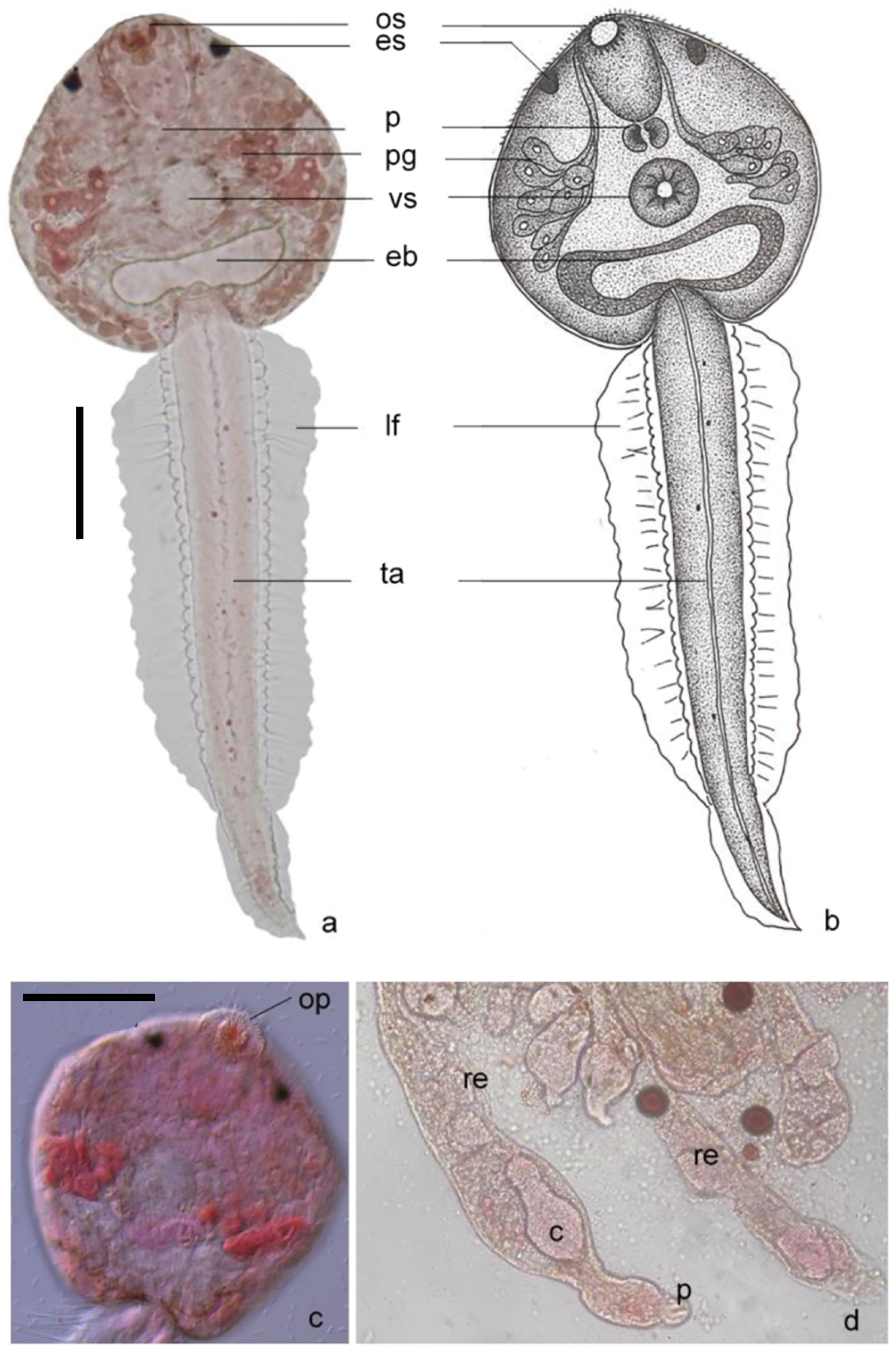
Image of Astiotrema monticellii Stossich, 1904 a. Images of cercaria stained with 0.5% neutral red (light microscopy) b. Drawing of cercaria structure c. Body part of cercaria stained with 0.5% neutral red (DIC microscopy) d. Images of rediae stained with 0.5% neutral red (light microscopy) Abbreviations: os: oral sucker, es: eyespot, p: pharynx, pg: penetration gland, eb: excretory bladder, vs: ventral sucker, ta: tail, lf: lateral finfold, op: oral spine, c: cercaria, re: redia. (Scale bars: 100 µm). |