|
||
|
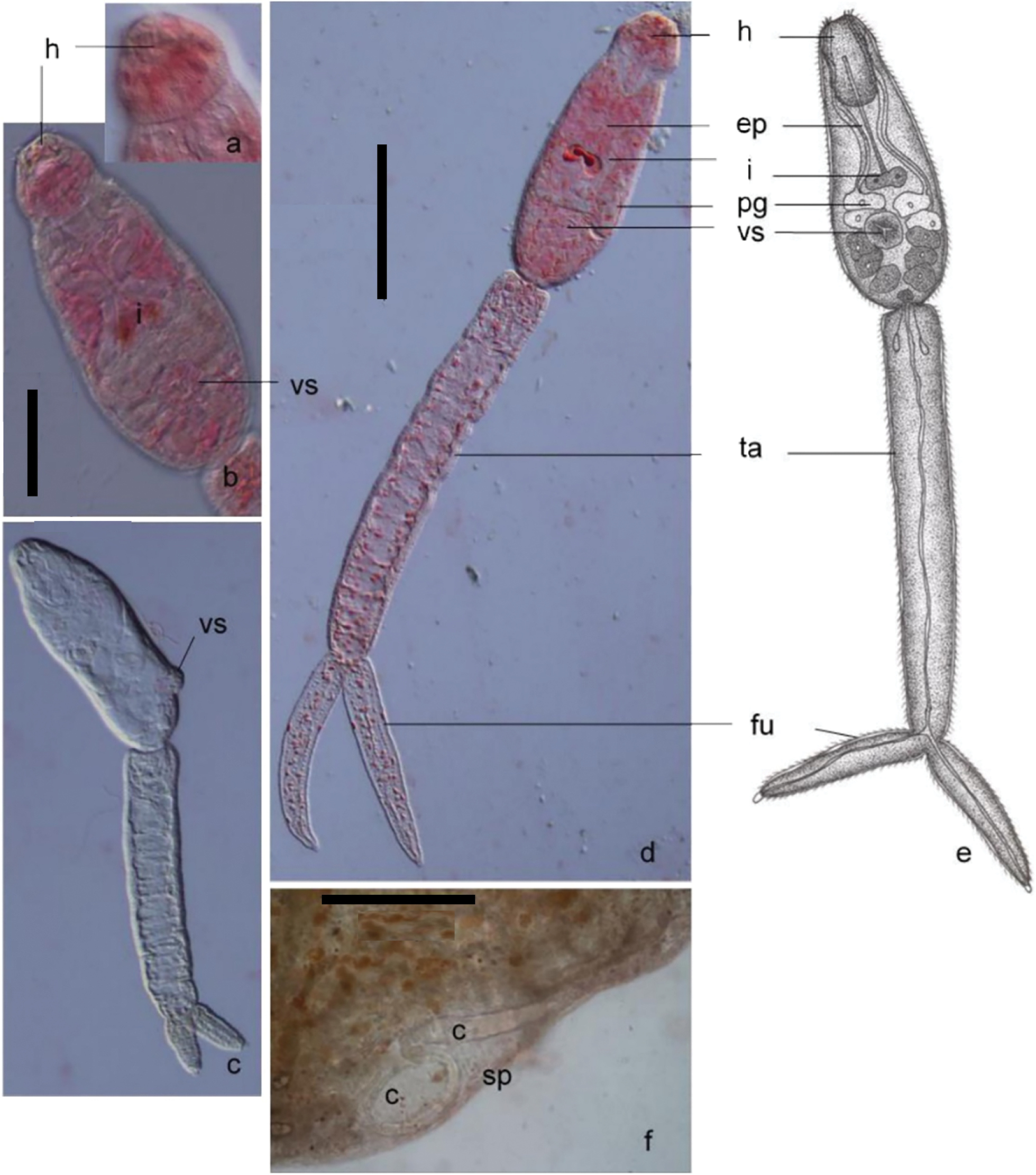
Image of Schistosoma indicum Montgomery, 1906 (Syn. S. nasalis Rao, 1933) a. Head organ of cercaria stained with 0.5% neutral red (DIC microscopy) b. Body part of cercaria stained with 0.5% neutral red (DIC microscopy) c. Image of unstained cercaria (DIC microscopy) d. Images of cercaria stained with 0.5% neutral red (DIC microscopy) e. Drawing of cercaria structure f. Images of sporocyst stained with 0.5% neutral red (light microscopy) Abbreviations: c: cercaria, eb: excretory bladder, ep: esophagus, fu: furca, h: head organ, i: intestine, pg: penetration gland, sp: sporocyst, ta: tail, vs: ventral sucker.. (Scale bars: 100 µm). |